For most cancers, the diagnosis is confirmed the same way, a pathologist examines the tissue itself.
What happened
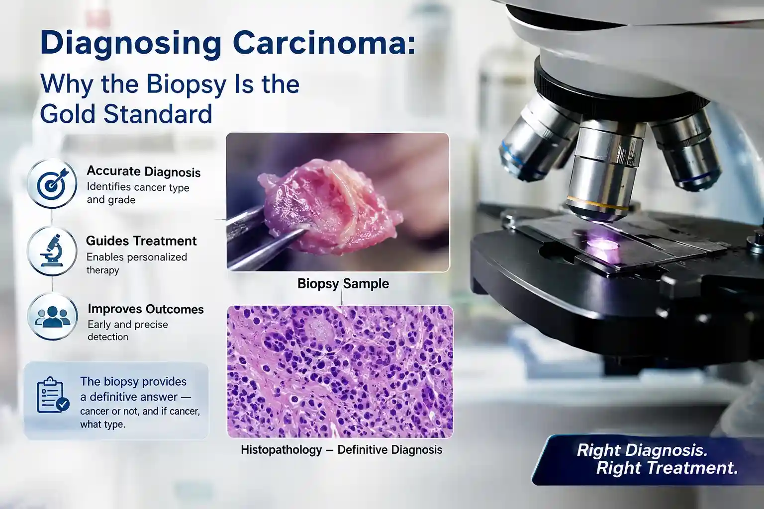
In 1858, Dr Rudolf Virchow, often called the father of cellular pathology, argued that disease begins in cells (“omnis cellula e cellula”, every cell from a cell). That idea put the microscope at the centre of diagnosis. A carcinoma is a cancer that arises from epithelial cells, the cells that line and cover the body’s surfaces and glands. Confirming one means examining a biopsy or resection under the microscope to see how the cells and tissue are organised.
Why it mattered
Imaging and blood tests can raise suspicion, but the biopsy remains the definitive answer. It shows whether a growth is benign or malignant, what type it is, and often how aggressive it looks (its “grade”). That diagnosis shapes the entire treatment plan. On difficult cases, an expert second-opinion review adds another layer of confidence before major decisions are made.
Key points
- A carcinoma is a cancer that arises from epithelial cells
- Histopathology of a biopsy or resection confirms the diagnosis and the tumour type
- Grading describes how abnormal the cells look and helps guide treatment
- Expert review / second opinion supports confident reporting on difficult cases
How this connects to our testing
This is the core of PathMole’s work, histopathology on biopsies from small samples to major resections, and expert second opinions. See the Test List, or contact the lab to discuss sample requirements. Pricing is shared on enquiry.
Sources
Virchow R: Cellular Pathology, 1858, establishing the cell as the basis of disease. · World Health Organization: Classification of Tumours (the standard framework for naming and grading tumours). See also our explainer on the WHO Classification of Tumours.